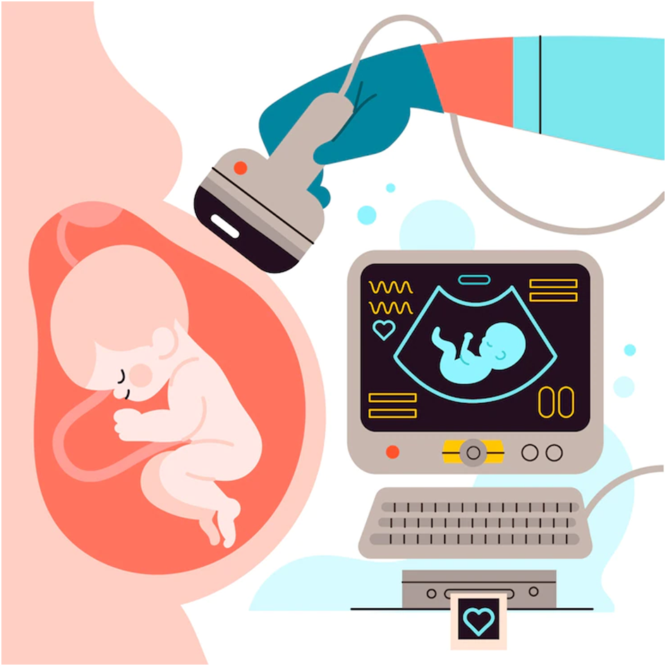
Your health can be learned a lot from ultrasound scans. We can examine various parts of your body with various types of ultrasound or even look inside a pregnant woman to see a developing baby.
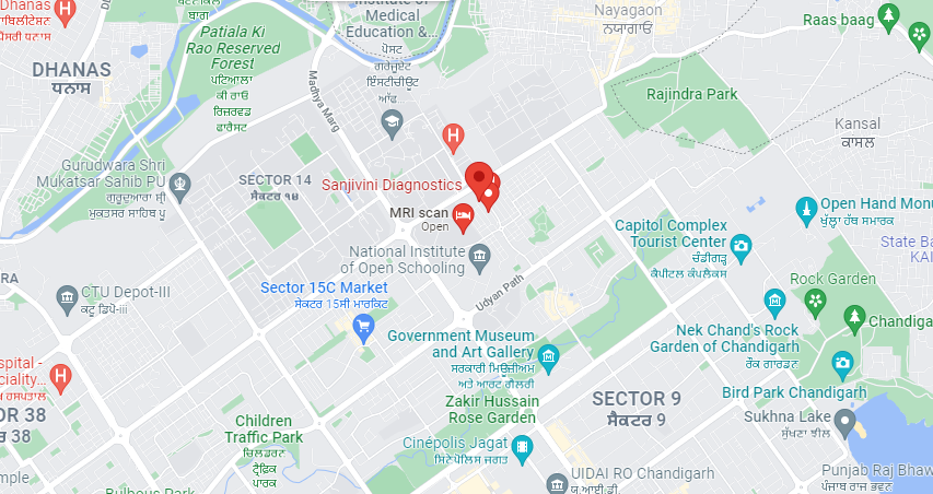
All of these various scans can be completed by Sanjivini Diagnostics at our Best ultrasound in Chandigarh clinic.
How does ultrasound work?
A particular kind of soundwave that is audible to humans is called ultrasound.
These soundwaves can be used by the technology in our private ultrasound clinic to produce precise images of your internal organs.
Different types of tissue inside your body will reflect the sound waves that are emitted by the ultrasound probe.
The probe will gather the ultrasound echoes and use them to create precise, real-time images of your organs that move.
Abdominal Ultrasound
Examining internal organs like the liver, gallbladder, spleen, pancreas, kidneys, and bladder using an abdominal ultrasound is helpful.
This can assist in the diagnosis of a number of ailments and the evaluation of the harm brought on by the diseases.
Ultrasound can also be used for the following because it produces real-time images:
- Describe techniques like needle biopsies, in which cells are taken from organs with needles and tested in a lab.
- Identifying the cause of many abdominal pains, such as kidney or gallbladder stones, can help a doctor treat the patient.
- aid in determining the reason for an abdominal organ’s enlargement.
- Major blood vessels are examined using a special type of ultrasound study called Doppler ultrasound.
- These images can aid the doctor in seeing and assessing:
clots and other blood flow obstructions. - plaque accumulation inside the vessel.
- a congenital defect.
Carotid & Abdominal Aorta Ultrasound Imaging
A quick, noninvasive way to find blood flow obstructions in the neck arteries leading to the brain that could cause a stroke or mini-stroke is with carotid ultrasound.
An aneurysm is an abnormal enlargement of the abdominal aorta that typically results from atherosclerotic disease, and it can be detected using an ultrasound of the abdominal aorta.
The patient is lying on a movable, tilting examination table. The area to be examined is covered with a clear gel.
Since sound waves cannot travel through the air, the gel aids in the transducer’s secure contact with the skin and removes any air pockets.
After viewing the images on the monitor and taking “snapshots” as necessary, the sonographer or radiologist presses the transducer firmly against the skin and sweeps along the area of interest.
Transvaginal Ultrasound
The same as for a gynaecological exam, a woman must empty her bladder prior to a transvaginal ultrasound.
Along with lying face up on her back, she is also wearing stirrups. For this test, the ultrasound transducer must be inserted.
In comparison to the typical speculum used in Pap tests, the transducer is smaller.
Before being inserted into the vagina, the transducer is covered with a protective covering and lubricating gel.
The transducer is only inserted into the vagina for the first two to three inches.
To obtain images from various angles, the doctor may move the device around.
Searching for the source of pelvic pain is the most frequent reason for transvaginal pelvic ultrasounds.
Conclusion
In order to measure the flow of blood through your arteries, we can also conduct a special kind of scan called a Doppler scan.
The carotid artery, which supplies the brain with blood, is typically the target of the scan. It can indicate your risk of developing a stroke, which frequently occurs when an artery becomes congested with fatty material.
Therefore, scans can aid in preventing potentially serious health issues.
You can schedule an appointment at Best Ultrasound in Chandigarh if you need to arrange any type of ultrasound scan in Chandigarh.